
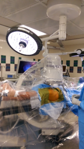
Surgeon and all instruments on the ipsilateral side. Arthroscopy cart on the opposite side at the patient’s shoulder level. C arm comes from the opposite over both legs at a 45 degree angle. C arm monitor is placed at the foot end.
For obese patients, a really long hip scope is available. A laparoscope with a long plastic cannula can also be used. The other hip instruments ae long enough and can slide on a slotted cannula. The arthroscope can be pushed into the fat to reach deeper, but that strains your shoulders by the end of the case. In rare situations, the incision can be enlarged to advance the bridge portion in.
For the central compartment first approach, which is most efficient and common in my practice, the hip is distracted in sight flexion, full adduction against a large perineal post, and internally rotated.
For peripheral compartment first, the hip is kept in 20 degrees of abduction and 30-40 degrees of flexion.
There are three ways to enter the hip joint arthroscopically.
1. Central compartment first
2. Peripheral compartment first especially when distraction or visualization are inadequate
3. Extra capsular endoscopy and capsulotomy first when peripheral compartment is not accessible due to capsular ossification.
Fastest way is to distract the joint and make either anterolateral or posterolateral portals under guidance. Even with the best technique, there is a small risk of labral or head damage.
My distraction technique
Paralyze hip muscles completely. Pad and secure both feet in foot holders on a fracture table. Operative hip in 20-30 abduction and opposite leg in neutral. Use a 8-10 inch perineal post. Place the table in 20-30 degree Trendelenburg tilt. Apply counter traction to the nonop leg to maintain the pelvis stable. Apply moderate traction to the operative hip. Adduct the hip against the post to produce lateral subluxation. Need not get final distraction of 7-10 mm at this stage. Release traction and prepare the hip. Reapply traction and insert spinal needle into the joint and inject air to get more distraction. Internally rotate the hip maximally to increase the anterior space.
Portal placement
Determine anterolateral portal path based on joint space and not based on trochanteric height. Insert needle at the anterior border of the trochanter. Aim posteriorly towards the lateral midline capsular entry closer to the head and the bevel of the needle away from the head. Check flouro shot when the needle touches the capsule. Enough space from the needle tip / lateral capsule to the air arthrogram showing the medial capsule suggests that the needle is close to the lateral midline and not too anterior or posterior. Maintaining the needle farther from the acetabulum and closer to the head decreases the risk of labral penetration. The needle entering the hip with a pop and advancing far to the medial acetabulum assures good position. Use the smallest diameter cannula with a blunt rounded tip trocar because damage will be less if portal is not well placed. Let the cannula bounce off the head if it hits the head. Try to make a second portal without putting fluid in.
In bloody situations, run fluid and suction a few times to clear the joint. Placing an outflow needle anywhere in the joint clears the picture quickly.
The choice of the second portal is based on the purpose and is always made under direct vision. If the anterior capsular triangle is wide, a standard anterior portal is the easiest. Good for synovectomy, loose bodies, labral debridement, neck osteoplasty. Bad for anchor placement in the acetabulum.
Distal anterolateral and mid anterior portals are more lateral, more difficult to place, and allow better trajectory for labral repair. Flouro can assist if triangulation alone is not successful.
Posterolateral portal is easy under direct vision or using flouro.
A wide capsulotomy makes everything easier with less traction force, but increases the risk of instability in dysplastic hips.
Do not get fixated on names, distances, or angles for portal placement. Anything lateral to the ASIS and Sciatic nerve is safe if you can reach where you want to reach.
Keep incisions small to prevent excessive fluid leakage. Use epinephrine to decrease pump pressure. Maintain lowest acceptable mean blood pressure.
I don’t distract the joint for acute septic arthritis. Single peripheral compartment portal is often adequate for irrigation and drain placement.
I go more medial when I am not able to make a more lateral portal, not the other way around.
I routinely do rim trimming, labral repair, and femoral neck osteoplasty through a standard anterolateral portal and a distal anterolateral (DALA) portal. Rarely, I may have to insert the 2 o clock anchor percutaneously and tie it through DALA.
Hip arthroscopy is not easy to learn. I learned it very slowly converting from open to arthroscopic techniques and had my share of complications. A hands on fellowship in a high volume center is the best way to learn, which is not practical for many. Next best is to read, watch techniques, attend cadaver courses, and visit experienced surgeons multiple times. AANA conducts 2 day courses 2-3 times a year and is a good start.