
Femoral neck osteotomy is best suited to correct the 3 dimensional deformity of slipped epiphysis that always has both translation and angulation in multiple planes at the head neck junction. It can be used to correct coronal plane deformities of the neck like Caput valgum, Coxa vara, and Coxa valga, but these can be corrected well with much simpler peritrochanteric osteotomies because they do not have any translation in the neck.
This osteotomy became safer when performed with surgical dislocation approach and extended retinacular flap development. This is the most difficult femoral procedure when performed for a severe fused SCFE. After the initial learning curve, Dr. Prasad Gourineni has good results with this procedure. It is different from the modified Dunn procedure performed for stable SCFE through the physis. Femoral neck osteotomy requires breaking the neck with an osteotome to separate the head that is fused to the back of the neck. In severe deformity, the posterior retinaculum cannot be elevated fully till the head is moved away from the greater trochanter. The osteotomy should be performed distal to the physeal scar, the metaphyseal bone left on the head needs to be removed, and then the head is reduced and fixed to the end of the neck. There are several potential complications and this should be attempted only after adequate training with it.
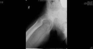
Femoral head fused to the back of the neck from a severe slip

Femoral neck osteotomy with complete correction of the slip