
Hip joint is a ball and socket joint designed for optimal balance between inherent bony stability and excellent functional range of motion. Variation from the normal morphology can lead to instability in the form of dysplasia or decreased range of motion in the form of femoro-acetabular impingement. Both can lead to premature osteoarthritis of the hip based on the severity of the abnormalities and the person’s activity level. Radiographic abnormalities of one part of the joint can be adequately compensated by a variation in another part of the joint and avoid arthritis. Occasionally the hip can show signs of both dysplasia and impingement at the same time.
Standard radiograph of the hip
Antero-posterior view
Evaluation of the entire pelvis is necessary in evaluating either hip joint, both for individual analysis of a hip joint as well as for comparison to the opposite side.
A standard pelvis x-ray is performed in supine position and the pelvic tilt and rotation are controlled to obtain similar x-rays in every patient. The functional variability of the hip position in standing has not been studied.
Neutral rotation
A good pelvis AP view should not have any rotational malposition. The central sacral line and the tip of the coccyx should align with the symphysis pubis. The two obturator foramina should appear symmetrical.
Neutral tilt
A good pelvis AP view should have neutral tilt and be neither close to an inlet nor an outlet view. The distance between the sacro-coccygeal junction should be 2-3 cm above the superior end of the symphysis in males and between 2-6 cm in females.
The exposure of the film should delineate the acetabular walls well. If the distal femoral rotation is controlled by keeping the patella perfectly anterior, the femoral version will be reflected on the film and the apparent neck-shaft angle may be an over estimate of the true angle. If the hips are held in internal rotation to bring both the trochanters parallel to the floor, the neck-shaft angle measurement will be more accurate.
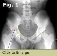
Figure 1. AP pelvis showing sourcil, sourcil angle, weight bearing axis, fovea and tear drop on the right hip, and posterior wall sign and cross over sign on the left hip.
Lateral view of the hip
False profile lateral- shows anterior acetabular coverage of the femoral head. Allows measurement of anterior center-edge angle and shows anterior subluxation during weight bearing. Only one hip can be studied at a time. A good AP pelvis will show the anterior coverage well and a false profile view may not be necessary.
Frog lateral – Both hips are held in flexion and maximal abduction and external rotation. The pelvic tilt is not controlled and the acetabulae cannot be analyzed well. As both hips are visible, the range of motion of the two hips can be compared. In hips with good motion, the visible part of the anterior femoral head-neck junction may be antero-inferior and may miss the cam deformity.
Shoot-through lateral / Long neck lateral in 15* of internal rotation – shows only one hip, but shows the cam deformity better and gives clues to femoral version.
Dunn lateral in 45* of flexion, maximal abduction, and neutral rotation – shows both hips for comparison, shows the antero-superior head-neck junction well, and gives a good estimate of femoral version.
Acetabular parameters
TearDrop
Teardrop is a radiographic condensation of the innominate bone at the inferior end of the acetabulum. A normal teardrop is U shaped. The medial border of the teardrop is continuous with the ilio-ischial line (a.k.a. Kohler’s line) and the lateral wall is continuous superiorly with the floor of the acetabulum.
The width of the teardrop varies with rotation of the pelvis. A wide teardrop is associated with a shallow acetabulum. A very narrow tear drop where the medial and lateral wall touch each other at the floor of the acetabulum or cross over is a sign of a deeper than normal acetabulum causing over coverage of the head called coxa profunda.
Sourcil
It is the radio-dense subchondral bone of the weight-bearing dome of the acetabulum. The lateral edge of the sourcil should be differentiated from any extra-articular ilium that gives a false estimate of head coverage.
Size – The sourcil normally extends laterally to cover about 80% of the width of the femoral head. The percent of head covered is difficult to measure if the head has a cam deformity. This coverage can also be measured by the anterior center-edge angle of Wiberg (normal 25-30*).
Slope – Sourcil also has a slope that can be measured. A straight line drawn from the medial edge of the sourcil to its lateral edge should be horizontal or up to 10* superior to the line connecting the inferior edges of the two sourcils (Sourcil or Tonnis angle). This maintains the joint reaction force transmitted along the primary compression trabeculae of the femur to be perpendicular to the slope of the sourcil. This normal alignment minimizes any shear stresses between the femoral head and the acetabular roof represented by the sourcil.
An increase in the up slope of the sourcil induces lateral subluxation of the femoral head, which will be resisted by the labrum and the capsule initially. These structures react with hypertrophy initially, but ultimately the labrum fails with degeneration and allows lateral subluxation of the head in dysplasia.
A down sloping sourcil induces medial translation of the head and loading of the acetabular fossa and fovea of the head. This abnormal loading results in medial osteoarthritis of the hip with well maintained superior joint space.
Version
The normal acetabulum is said to be anteverted by 20 degrees. The version of the acetabulum is hard to measure in any cross sectional study as it varies by the distance of the cut from the dome. A global estimate of the version is best noted on the AP view.
The anterior wall of the acetabulum is always more horizontal and extends towards the pubis. The posterior wall is more vertical and extends to the ischium, which is lateral to the pubis. In the normal anteverted acetabulum the anterior and posterior walls contact each other at the lateral edge of the sourcil and should not cross each other.
The anterior wall covers the head to a less extent than the posterior wall, and the posterior wall typically passes through the center of the head. If the posterior wall passes lateral to the center of the head, the posterior coverage is considered to be excessive and if it passes medial to the center of the head (posterior wall sign), the posterior coverage is deficient.
A very medial anterior wall that barely covers the femoral head as in dysplasia suggests deficiency of anterior coverage. Over coverage posteriorly and under coverage anteriorly should be considered as excessive anteversion of the acetabulum. Anterior over coverage is suggested by any crossover of the anterior wall over a normal posterior wall, while true retroversion of the acetabulum should have posterior under coverage along with the crossover sign. The lower the cross over occurs the more significant is the anterior over coverage.
Appearance of the ischial spine on a good AP view is a sign of acetabular retroversion.
Subchondral cysts can develop from rim loading in dysplasia and after advanced damage from cam impingement.
Osteophytes of the rim of the acetabulum can develop from ossified labrum from pincer impingement.
Os acetabulare and rim fractures can occur from rim loading in dysplasia and from impingement.
Femoral parameters
Shape of the head
The femoral head is close to a sphere. Loss of sphericity by flattening, or overgrowth of the epiphysis on to the neck produces a misshapen head that may not be congruous with in the acetabulum. This sphericity may be measured objectively using Mose circles.
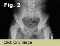
Sagging rope sign on the AP view shows the abnormal extension of the head on to the neck. (Blue line in Fig 2)
Figure 2 – Cam Impingement
Coxa magna (large head) is not problematic if it is well contained and is congruous in the acetabulum.
Physeal scar
The location of the physis can be seen even after closure of the physis. Superolateral extension of the physeal scar with extension of the epiphysis on to the superior neck can be seen on the AP view (orange line in Fig 2). This is typical of the cam deformity of the head, which appears like a bicycle helmet on the lateral view performed at the appropriate rotation.
Fovea centralis
Fovea is a depression in the femoral head for the attachment of ligamentum teres. Fovea is located medially and inferiorly on the head and does not come in contact with the sourcil. The articular cartilage inferior to the fovea is thinner and has less surface area compared to the cartilage superior to the fovea.
A superiorly placed fovea coming in contact with the sourcil is abnormal and is called Caput valgum or Fovea alta.
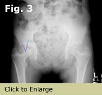
Figure 3 – Caput valgum
Effective articular surface
The area of contact between the articular cartilage of the head and the sourcil can be measured by an angle between a line extending from the center of the head to the upper edge of the fovea or the medial edge of the sourcil (whichever is more superior) and a line from the center of the head to the lateral edge of the sourcil. This area should be large without causing impingement. top of page
Femoral neck
The function of the femoral neck is to provide a lever arm for the abductor muscles and to provide adequate clearance around the head for normal range of motion.
Neck-shaft angle The angle between the femoral shaft axis and the femoral neck axis is usually between 125-130*. Internally rotating the hips till the neck is horizontal to the floor shows the true angle while any external rotation of the femur will increase this value. The axis of the neck is difficult to define and the neck shaft angle can be changed by surgically removing the superior aspect of the neck without changing the shaft and neck relationship.
Coxa valga increases the resting length of the abductors but decreases the abductor lever arm and results in increased joint reaction forces.
Coxa vara decreases the resting length of the abductor muscles, causes abductor fatigue, Trendelenburg gait, decreases the joint reaction forces in the hip by increasing the abductor lever arm.
Coxa breva (short neck) decreases abductor resting length and lever arm, increase joint reaction forces, and causes abductor fatigue and Trendelenburg gait.
Trochanteric height – The tip of the trochanter lies at the level of the center of the femoral head. In coxa vara, it is superior to the center of the head and in coxa valga; it is inferior to the center of the head. This relation is minimally effected by any rotation of the hips. top of page
Version
Version of the femur is the angle between the femoral condylar axis and the femoral head neck axis. Normal version of the femur is around 20* of anteversion or internal torsion of the condyles in relation to the femoral head and neck axis. The femoral neck axis represents the head neck axis usually except; with slipped capital femoral epiphysis the head tilt deformity changes this axis.
Lateral offset / Articulotrochanteric distance
The distance between the center of the head and the tip of the trochanter on the horizontal plane shows the abductor lever arm. It is normally 2 times the diameter of the head.
The lateral offset is inversely proportional to the joint reaction forces in the hip.
Head-neck offset – Alpha angle
A narrow neck relative to a larger head is desirable to maintain good range of motion. In artificial femoral components the diameter of the head and the diameter of the neck are precisely determined to calculate the head neck offset, such measurements are less useful in the native hip due to circumferential variability of the head and neck offset.
A narrow neck around a spherical head will allow motion till the acetabular labrum contacts the femoral neck at it’s maximal concavity. The most common area of impingement is at the anterior femoral neck and a less common area is the posterior neck. Anterior impingement occurs at less flexion when the abnormal head-neck offset is more superior on the anterior aspect of the neck.
Abnormal offset is noted on the lateral view when the head goes out of sphericity more medial than normal. Alpha angle is the angle between the neck axis and the line connecting the center of the head to the point at which head sphericity ends. The normal alpha angle is about 45* and a higher angle implies less motion before impingement occurs.
Figure 4 – Increased alpha angle (70*) from cam deformity.
Herniation pit occurs at the site of impingement on the femoral neck.
Congruity of the hip
The articular surfaces of the femoral head and the acetabulum are usually parallel to each other at the dome of the acetabulum. Lateral subluxation of the head causes the space to be wider medially. Mild lateral widening is seen in asymptomatic hips though it usually denotes medial subluxation.
Medial space
Distance between femoral head and Kohler’s line is normally between 10-15 mm. Increased space is seen in dysplasia and lateral subluxation. Decreased space is seen with coxa profunda and the femoral head touches or crosses the Kohler’s line in true protrusio acetabulae.
Shenton’s line indicates the continuation of the superior obturator foramen with the inferior femoral head and neck. Superior and lateral subluxation is suggested by a break in the Shenton’s line as the head and neck of the femur lie superior to the superior border of the obturator foramen. Excessive external rotation of a normal hip can break the Shenton’s line without any subluxation of the head.
Summary
|
|
Dysplasia |
Normal |
Anterior FAI |
|
Teardrop |
Wide |
narrow ‘U’ |
crossover |
|
Sourcil |
|
|
|
|
% Coverage |
<70% |
80% |
>90% |
|
CE angle |
<20* |
25-30* |
>35* |
|
Sourcil angle |
>10* |
0-10* |
<0* |
|
Acetabulum Version |
^anteversion |
normal |
retroversion |
|
Anterior wall |
deficient |
normal |
crossover |
|
Posterior wall |
nl / ^ |
normal |
nl / deficient |
|
Femur Version |
> 30* |
20* |
<10* |
|
McKibbin Index (Combined anteversion) |
>40* |
30-40* |
<30* |
|
Fovea |
alta |
normal |
normal |
|
Neck-shaft angle |
valgus |
normal |
varus |
|
Alpha angle |
45* |
45* |
>50* |
|
Medial space |
>15mm |
10-15mm |
10-15mm |
|
Shenton’s line |
disrupted |
intact |
intact |
*The numbers above are basic differentiators of individual hip parameters. Occasionally, a hip can have certain parameters of FAI and others suggesting dysplasia making clinical exams important.